Archive
Structural studies of naphthalene diimide ligands with telomeric G-Quadruplex DNA
Structural Basis for Telomeric G-Quadruplex Targeting by Naphthalene Diimide Ligands
Gavin W. Collie, Rossella Promontorio, Sonja M. Hampel, Marialuisa Micco, Stephen Neidle*, and Gary N. Parkinson*
J. Am. Chem. Soc., 2012, 134 (5), 2723; DOI: 10.1021/ja2102423
A synopsis by Maxier Acosta
Previously Neidle had reported a series on naphthalene diimide (ND) oligo G-quadruplex (OGQ) ligands with side-chains (n) of 3-5 carbons with N-methyl-piperazine end groups. They showed experimentally how it inhibited binding of hPOT1 and topoisomerase IIIα to telomeric DNA and inhibited telomerase activity in MCF7 cells via the stabilization of OGQs (DOI: 10.1016/j.bmcl.2010.09.066). Now, in collaboration with Parkinson, they report the crystalline structure of each one of those naphthalene ligands with the addition of a two-carbons side-chain.
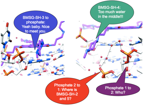
They first give an overview of the tendencies of the overall parallel OGQ (Gtel22) with each ND ligand. With the telomere sequence d(AGGG[TTAGGG]3) they highlight the stacking of two OGQs making a dimer interacting from the 5’ terminal G-quartet. But the ratio between the ND and each OGQ is 1:1. Taking this in consideration, when each ND is bound to the quadruplexes, they force the topology of the loops into parallel strands as first proposed in DOI: 10.1016/j.bmcl.2010.09.066. While going more into detail, stability studies via FRET and inhibition studies where done for each ND. In the case of the ND with a two-carbons side-chain, it didn’t enhanced by much the stability of the Gtel22 due to the inappropriate side-chain length to enable effective interactions (in the OGQ groove) between the protonated N-methyl-piperazine and the DNA backbone phosphates. Although the n=5 ND OGQ complex showed poor quality in its crystal diffraction, it was still higher than that corresponding to n=2. For the n=4 ND, the side-chains were too long to fit well into the grooves as indicated by the disorder of the chains leading to a decrease of strong specific contacts, yet it was still more stabilizing than n=5 ND. For n=3 ND, it was observed that the cation-phosphate interactions were specifically coordinated, making it the best ligand of the small library presented in the paper. The structural features for these ND ligands correlated well with the inhibition of two types of cancer cells (MCF7 and A549).
In the discussion they summarized the data in three major topics: (1) the 1:1 binding of ND and OGQs; (2) the importance of the electrostatic side-chain interaction with the groove; and (3) the retention of the parallel topology of the Gtel22. Also, as might be expected for scientists from a pharmacy school they maintain their focus on how biologically relevant these binders could be for anticancer treatments.
In general, I thought that this was a good OGQ-binder structural article. I know that our systems are difficult to crystallize, yet this type of studies can help us to understand them to a new level so we could also start talking about potential inhibitors among other things. In terms of the organization of the paper, I found confusing the fact that they do not address explicitly some of the figures. In the discussion it was not that clear for me why the NDs induced the parallel topology; so, for that I encourage you guys to read the reference that I mentioned at the beginning, which has additional useful experimental data that may help anyone in the same situation. Other than this, I wish I had seen all of the ND side-chains interactions with the groove (some of them are in the supplementary information).
Raiders of the lost G-quadruplexes …in the human genome
Small-molecule–induced DNA damage identifies alternative DNA structures in human genes
Raphaël Rodriguez, Kyle M Miller, Josep V Forment, Charles R Bradshaw, Mehran Nikan, Sébastien Britton, Tobias Oelschlaegel, Blerta Xhemalce, Shankar Balasubramanian* & Stephen P. Jackson*
Nature Chemical Biology 8, 301–310 (2012) doi: 10.1038/nchembio.780
A synopsis by Diana Silva Brenes
The authors of this week’s paper play detective to find out -with great detail- what exactly happens to a human cell when it’s treated with the versatile, potent GQ-binder, pyridostatin. Using a combination of biomolecular assays, the authors manage to give strong support for the in vivo formation of GQ-DNA in human cells, and show their role in the activity of the new drug.
Pyridostatin is shown to induce damage to cellular DNA, stumping their proliferation. This happens because cellular checkpoints, which revise DNA before continuing the cellular division cycle, detect the damage and signal to the cell that something is wrong. The cell stops in its tracks to try to correct the problem before it continues the cycle. The drug, however, isn’t too toxic and most cells can survive long-term exposition to it without undergoing apoptosis. Interestingly, inhibition of the checkpoints restores cell proliferation.
Many of the results rely on detecting the presence of γH2AX (a protein that indicates double strand breaks in DNA) as a way to follow damage done to DNA. In cells treated with pyridostatin, γH2AX is present during the DNA transcription and replication processes, pointing at damage to DNA occurring during both stages.
Next, the authors wanted to localize where in the DNA is pyridostatin taking effect. Fluorescence labeling of γH2AX and the telomeres (marked by the labeling of a telomere binding protein) didn’t show co-localization. It was, thus, necessary to modify the drug to add direct fluorescence labeling. Addition of an alkyne group to the drug allowed an in cellulo click reaction with an azide containing fluorescent dye. After making sure that the modified pyridostatin did not affect drug activity, staining of pyridostatin was performed and fluorescent spots (foci) were compared with the a fluorescently labeled human helicase reputed to bind and resolve GQ-DNA during replication. Good co-localization was observed, suggesting that pyridostatin was localized mostly at putative GQ-DNA sites. In another experiment they showed that addition of pyridostatin before of after “freezing” the cellular processes in formaldehyde gave almost identical results, suggesting that GQ structures are pre-folded even without addition of pyridostatin.
They then performed ChIP sequencing to try to figure out which genes (aka, DNA segment) were targeted by pyridostatin. They found several specific genes (mostly away from the telomeres) that sustained pyridostatin induced damage to DNA, and all of them had above average putative GQ sequences. However, not all areas enriched in putative GQ sequences were affected, suggesting that there are other important requirements for interaction.
A particularly affected gene was SRC as confirmed by checking for loss of its corresponding mRNA transcription activity. Out of 25 putative GQ sequences estimated for this gene, 23 of them could be observed to form QGs in vitro using CD and NMR spectra.
The effect of pyridostatin on the bioactivity of SRC was also evaluated. SRC is important for wound healing and motility of cells. Cells treated with pyridostatin displayed a reduced ability to heal. As a control, cells treated with another DNA-damaging drug (DOX), didn’t affect healing, proving that the deficiency was not due merely to DNA damage.
It was previously shown that pyridostatin binds to GQs with enough strength to resist polymerases. It is hypothesized that damage to DNA by pyridostatin is due to mechanical forces breaking the DNA during the cell’s attempt to transcribe or replicate DNA. The findings of this paper support the potential drugability of GQs in cells.
The data reported by this paper is really important for the field of GQ binders and raises large hopes for the future of the field. Being able to use GQ to recognize and regulate specific genes is a dream come true in drug design, and the authors present strong data as to the viability of this approach. As a chemist, it’s difficult to get used to the rather indirect type of evidence that supports these findings, making it hard for me to comment on this paper’s methods. However, the controls and the analyses they did appear to be adequate. Overall, I find the results in this paper to be really important to anyone in the GQ field.
Battle for supremacy between G-Quadruplex DNA fluorescent probes
Fluorescence properties of 8-(2-pyridyl)guanine “2PyG” as compared to 2-aminopurine in DNA
Anälle Dumas and Nathan W. Luedtke*
ChemBioChem 2011, 12, 2044–2051; DOI: 10.1002/cbic.201100214
A synopsis by María Del C. Rivera-Sánchez
The motivation of the work reported by Dumas and Luedtke is the development of internal probes for direct readouts of local nucleobases arrangements, dynamics and electronic properties (e.g., electron transfer reactions). Their strategy is based on the incorporation of internal fluorescent probes as energy acceptors in DNA, particularly in hTelo and cKit sequences that fold into oligo-G-quadruplexes (OGQs).
In this article the authors include many of their previously reported data related to 2PyG [Refs 17 and 18] in order to compare its properties with those of 2-aminopurine (2AP), a nucleoside that was not previously evaluated as an internal fluorescent probe for OGQs when directly incorporated into folded G-tetrads. Each publication has different pieces of the puzzle towards understanding the importance of 2PyG as a plausible fluorescent probe and how it compares with other potential probes like 2AP. Thus, from those “scattered” pieces of information the picture that emerges can be summarized in the synthesis of a small family of 8-substituted-2’-deoxyguanosine analogues (2PyG, 4PVG and STG) and the evaluation of their photophysical properties in CH3CN and H2O. The cool part is that the phosphoramidite versions of these analogues were synthesized and the nucleosides incorporated into strategic positions of hTelo and cKit OGQs. The impact on the global structure and stability of hTeloG9, hTelo17, hTeloG23, cKitG10 or cKit15 having 8-substituted analogues, 2AP or thymine directly incorporated into folded G-tetrads, was evaluated by means of circular dichroism (CD) and CD-melting assays. Experiments using the afore mentioned ss OGQs were done in K+-, Na+-, and Li+-buffer and were compared to data from ds hTeloG9, ds hTeloG17, ds hTeloG23, and ds cKitG15 in Na+-buffer. In addition, the proficiency of analogues like 2PyG, 2AP and thymine as internal fluorescence probes was assed by measuring the quantum yield (Φ) and energy-transfer efficiency (ηT) of the substituted-duplex and ss-OGQs.
The data gathered from these experiments points to 2PyG as an outstanding internal fluorescent probe due to its higher quantum yield (Φ), once incorporated into folded oligonucleotides (Φ = 0.03–0.15) versus the free nucleoside in water (Φ =0.02), when compared to all other nucleosides evaluated. In addition, when exciting at 260 nm, the energy-transfer efficiencies from unmodified bases to 2PyG are 4–10-fold higher in ss-OGQs than in the corresponding duplex DNA. This energy-transfer process is favored by the O6 ion coordination within the central channel of G-tetrads and is distinctive of GQ structures (not duplex DNA). When this phenomena is combined with the high molar absorptivity of DNA it results in fluorescence enhancements of 10–30-fold for 2PyG-containing OGQs versus the corresponding ss- or ds-DNA. This highlights the potential of using 2PyG as a fluorescent probe for the detection of OGQ formation at lower concentrations among other applications. Unfortunately, the Φ or ηT of 2AP-containing DNAs are much lower than those for 2PyG-containing DNAs.
The ideal internal fluorescent probe should have very little effect on the global structure of the system evaluated. Particularly, the effect of 2PyG incorporation within folded G-tetrads seems to be context dependent. For example, 2PyG have little impact on the global structure and positive stability of hTeloG9 in K+- or Na+-buffer do to the syn conformational preference shared by this position and 2PyG. However, even though G15 in cKit (wt) have an anti conformational preference, CD spectra suggest that the incorporation of 2PyG have little impact on the global structure, but caused a small decreased in the Tm of cKitG15. On the contrary, the incorporation of 2PyG at hTeloG23 (in K+ or Na+-buffer) just allows the formation of an OGQ structure where G23 is in a syn conformation that is mainly observed on Na+-buffer. As a general trend, considering all the data discuss in the article, we can say that base stacking and pairing interactions can sometimes overcome the energy barrier of a preferred glycosidic bond conformation stabilizing the resulting OGQ or ds-DNA structure. Still, 2PyG has to be strategically located within OGQs to minimize detrimental effects, although, similar substitutions with 2AP or thymine are much more significant. Regarding 2AP, a priori I would not consider it a good mimic of guanine when positioned directly into folded G-tetrads because it lacks a carbonyl at the C6 and the N1-H, which prevents the formation of at least three interactions essential for an effective participation in the formation of a G-tetrad. Therefore, I consider that the comparison of 2PyG against other 8-substitutted nucleobases as they did on ref. 18 is more appropriate than comparing it against 2AP. The system reported by Dumas and Luedtke might have applications on fundamental studies related to ODNs and/or OQGs dynamics and their electronic properties, but I don’t picture them into practical, biophysical or technological applications.
This was a nice article in which it the authors combined many previous results with new complementary data provides a better understanding of the true potential and limitations of 2PyG as an internal fluorescent probe. They also evaluated for the detrimental effect induced by 2AP when incorporated into folded G-tetrads. The experiments reported included the appropriate controls like those done using thymine-containing sequences. In addition, their experimental section includes appropriate details such as the preparation of the DNA samples used.


